Image Quality of Digital Direct Flat-Panel Mammography Versus an Indirect Small-Field CCD Technique Using a High-Contrast Phantom
Figure 1
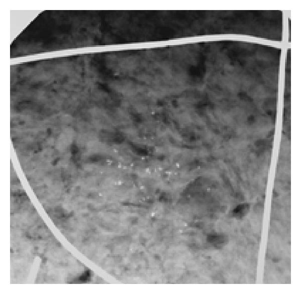
Radiological image of a laser-printer-film covered with different silica beads containing a polymethylmethacrylate (PMMA) sheet of 1.5-cm thickness (Plexiglas, Degussa) and a 1.5-com thick layer of ground meat as scattering bodies. For the raters’ orientation, a metal wire was used to divide the phantom into 4 quadrants. Direct flat-panel detector mammography scanned the whole phantom (a). The indirect CCD technique only produced spot images of the phantom’s 4 quadrants (c and d). Quadrant IV contains 49 lobular microcalcifications of 300–599 m in diameter.
(a) Direct flat-panel detector mammography. Survey image of the phantom
(b) Direct flat-panel detector mammography. Magnification of an area from (a)
(c) Spot film of quadrant IV using the indirect CCD technique with a 1024 acquisition matrix
(d) Spot film of quadrant IV using the indirect CCD technique with a 512 acquisition matrix